Quantitative evaluation of midbrain atrophy may be useful in differentiating progressive supranulear palsy psp from parkinson disease pd.
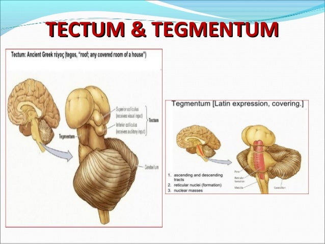
Roof of the midbrain.
Swelling on the side of the tectum responsible for vision.
The midbrain also known as the mesencephalon is the most superior of the three regions of the brainstem.
The tectum from latin for roof makes up the rear portion of the midbrain and is formed by two paired rounded swellings the superior and inferior colliculi.
The superior colliculus is a structure lying on the roof of the mammalian midbrain.
Midbrain the roof plate of the midbrain is formed by two paired rounded swellings the superior.
The superior colliculus is a layered structure with a number of layer.
The tectum latin for roof is the dorsal side of the midbrain.
It is a paired structure and together with the paired inferior colliculi forms the corpora quadrigemina.
The adjective form tectal is commonly used for both structures.
Mesencephala or mesencephalons is the most rostral part of the brainstem and sits above the pons and is adjoined rostrally to the thalamus during development the midbrain forms from the middle of three vesicles that arise from the neural tube.
The characteristic structures of this area are the superior and inferior colliculi.
In non mammalian vertebrates the homologous structure is known as the optic tectum or optic lobe.
It is involved in certain reflexes in response to visual or auditory stimuli.
Swelling on the side of tectum responsible for hearing.
It acts as a conduit between the forebrain above and the pons and cerebellum below.
In mammals the superior colliculus forms a major component of the midbrain.
Tectum the superior colliculus of the rostral area of the midbrain is part of the.
The superior colliculus receives input from the retina and the visual cortex and participates in a variety of visual reflexes particularly the tracking of objects in the visual field.
We determined whether an abnormal superior midbrain profile flat or concave aspect is a more practical diagnostic parameter.
However this finding is not specific of psp and quantitative measurements are not always practical.
The position of the tectum is contrasted with the tegmentum which refers to the region in front of the ventricular system or floor of the midbrain.
Intermediate level of midbrain.
Posterior to the cerebral aqueduct is the tectum roof of the midbrain figs.
In this article we will discuss the anatomy of the midbrain its external anatomy internal anatomy and vasculature.
Inferior and superior colliculi corpora quadrigemina.
In midbrain the tectum from latin for roof makes up the rear portion of the midbrain and is formed by two paired.
Covers several midbrain structures.
The midbrain is divisible into three regions which can be appreciated best in cross section.
The ventral portion of the midbrain is known as the.
When viewed in cross section the midbrain can be divided into three portions.
Structure of the brain in human nervous system.